RSS Google AI Blog
Follow
A new light on neural connections
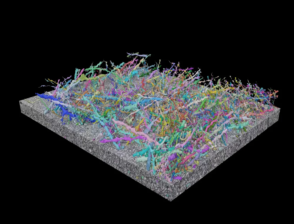
Antoine van Leeuwenhoek was the first person to document microscopic views of bacteria, red blood cells, and sperm cells using a homemade light microscope. Light microscopy has since become a fundamental technique in life science laboratories, but it has not been able to penetrate the field of connectomics. Connectomics is an area of neuroscience that has relied on electron microscopy, which requires expensive and highly specialized equipment. Researchers have now developed a method called LICONN that uses light microscopy to comprehensively map all the neurons and their connections in a block of mouse brain tissue. This was achieved by customizing several well-established techniques and combining them into a single workflow. The method involves physically expanding brain tissue while preserving structural integrity and chemically labeling all proteins to provide image contrast. The researchers validated LICONN by providing an automated reconstruction of a nearly one-million cubic micron volume of mouse cortex and demonstrating that it works comparably well to electron microscope-based connectomics. LICONN unlocks the ability to simultaneously measure structural and molecular information in a tissue sample, enabling fundamental new opportunities to understand the workings of the brain. The researchers are now working on scaling up LICONN to capture data from larger tissue volumes and are collaborating on projects to map a mouse brain and understand how brain structures change in the context of diseases like Alzheimer's.